The average life expectancy for both sexes increased by more than 50% from 1950 (47 years) to 2020 (73.2 years). There are lots of reasons behind this phenomenon, and one of the important reasons is modern medicine’s development. One particular technology that has to be mentioned is biomedical imaging, which breaks the traditional diagnostic strategy with its painless and noninvasive features to reduce patients’ fear. Because of the high sensitivity, high spatial resolution, quick diagnostic time, and real-time monitor, biomedical imaging can be used by physicians to diagnose diseased parts or even detect potential diseased parts as early as possible that can increase patients’ survival rates.
Magnetic Resonance Imaging (MRI), X-ray imaging, Computed Tomography (CT), and Optic imaging are the main detecting methods used in biomedical imaging. This article mainly focuses on X-ray imaging and Computed Tomography (CT) Scan. Let’s first get some basic knowledge about X-ray imaging and CT scan’s working principles.
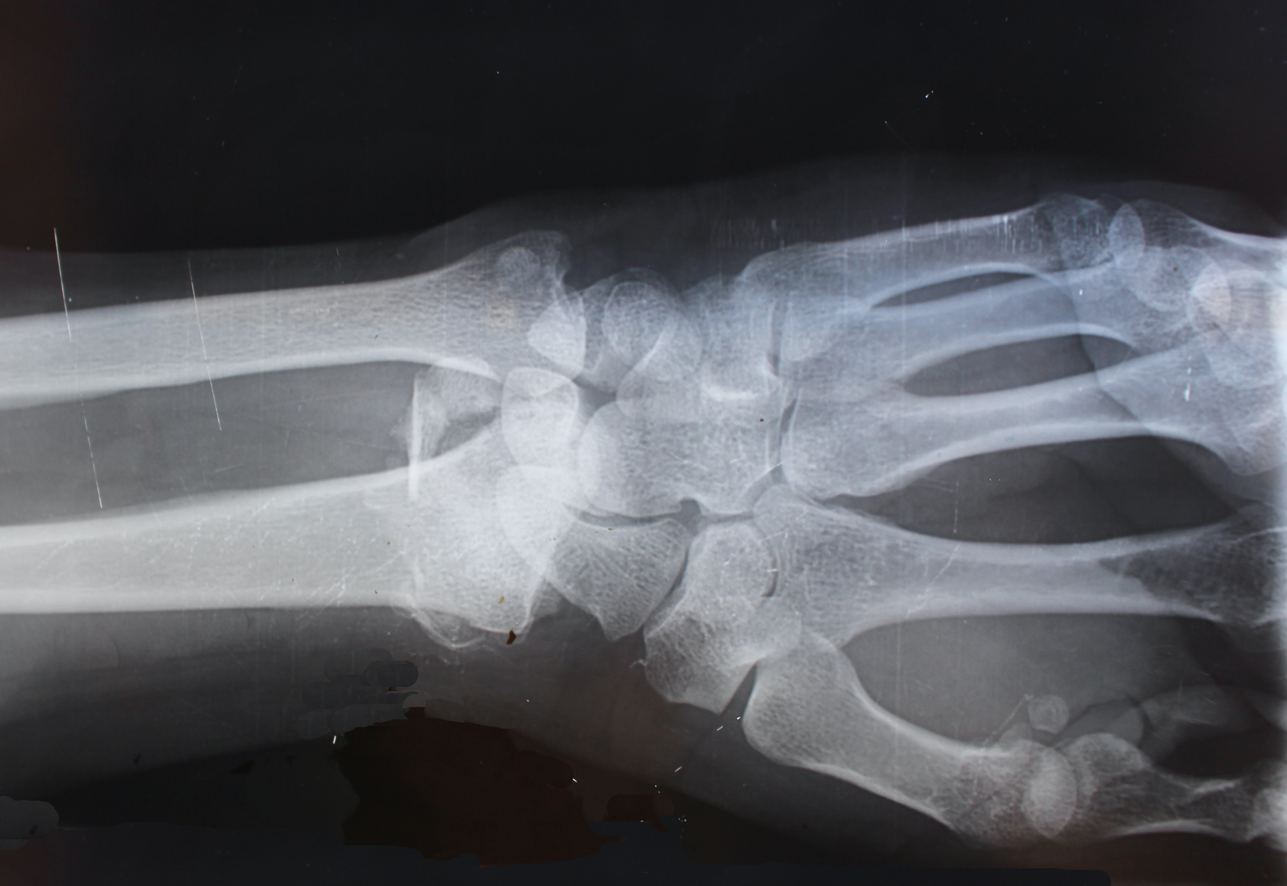
CT Scan is also called X-ray CT imaging. It is similar to X-ray imaging since they both use X-rays to “penetrate” a patient’s body. X-rays will be detected by an ionization chamber, an exposed film or digital imaging techniques.
When X-rays pass through the body, they can cause 3 mechanisms in the body tissues [1]:
The sum of these 3 mechanisms is described by the X-ray mass attenuation coefficient, which increases with increasing atomic number The X-ray attenuation coefficient shows the ability of a material to absorb or attenuate X-rays when they “penetrate” the material. And it can be calculated by the Beer-Lambert law.
I: the X-rays’ intensity after “penetrating” the material
I0: the X-ray’s initial intensity
μ: the X-ray attenuation coefficient
x: the material’s thickness
The larger X-ray attenuation coefficient μ is, the smaller “penetrating” X-rays’ intensity I is, which helps physicians to see body tissues more clearly. This allows denser body parts such as bones can be imaged clearly by a CT scan by increasing the X-ray attenuation coefficient.
As mentioned above, a detector will be in the back of the material to detect I value and the computer will transform these signals into usual X-ray images we often see. These are the basic working principles for X-ray imaging. CT scans use the same principles but additionally use a circular rotating X-ray source and detector that are controlled by a computer. Figure 1 represents a CT scan machine which can reconstruct a 3D image and keep a good contrast between soft tissues.
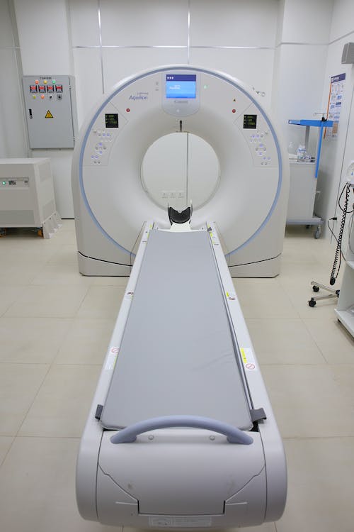
Figure 1: CT scan machine
Some parts of body tissues have similar density and atomic structure, which can’t be distinguished by X-ray CT. Contrast Agents (CAs) are created to help clinicians to increase the contrast of the interesting regions. Today most widely used X-ray CT CAs are iodine-based agents and barium sulfate. Due to iodine and barium’s high atomic number, iodine-based and barium-based agents have large X-ray attenuation coefficients.
Iodixanol is often used in arterial or coronal imaging. Iohexol or iopromide is used in brain and cerebral imaging. Ioxaglate is used in whole body imaging. Barium sulfate is often used in gastrointestinal tract imaging.
But these CAs can cause side effects to patients. About 30% patients bear painful, uncomfortable, or warm injections of intravenous iodixanol. Other phenomena such as high fever, low blood pressure, nausea, headache, and taste perversion are reported after using iodine-based agents [2]. Last but not least most iodine-based agents are not biodegradable and environmentally friendly [2].
A new type of CA is needed to change this situation. Rare-earth Nano Contrast Agents are developed to replace the general iodine-based CAs.
Due to rare-earth elements’ large atomic number (Z≥70) except scandium and yttrium, rare-earth nano contrast agents have large X-ray attenuation coefficients. They give better CT contrast images than clinical CAs such as iodine-based agents (Z=53) at the same concentration. Coated with organic molecules, rare-earth nano CAs can be more biocompatible than clinical CAs. Doping with other elements, mixing 2 or more nanoparticles, or combining them with different structures such as core shell give these nanoparticles more abilities to be used in multiple biomedical imaging such as CT, MRI, PAT, etc [2].
Gadolinium (Gd) and Ytterbium (Yb) nanoparticles are the 2 classical groups of rare-earth nano CAs. NaYbF4:Er doped with Gd3+ and coated with DSPE-PEG2000 shows very good biocompatibility and low cytotoxic effect [2]. NaLuF4:Gd nanoparticles can be used in dual-mode imaging: CT and up-converging imaging. Figure 2 shows that by using these nanoparticles, lung blood vessel imaging is improved greatly [2]. Gd2O2S:Eu3 doped with Yb nanoparticle can be used as a dual contrast agent for MRI and CT. NaYbF4 doped with Tm3+ can be used for CT and NIR dual bioimaging [2].
Rare-earth nano CAs show good biocompatibility and no toxic effect. By doping with different elements, combing different nanoparticles or even building different nanostructures, rare-earth nano CAs have countless possibilities for biomedical imaging.
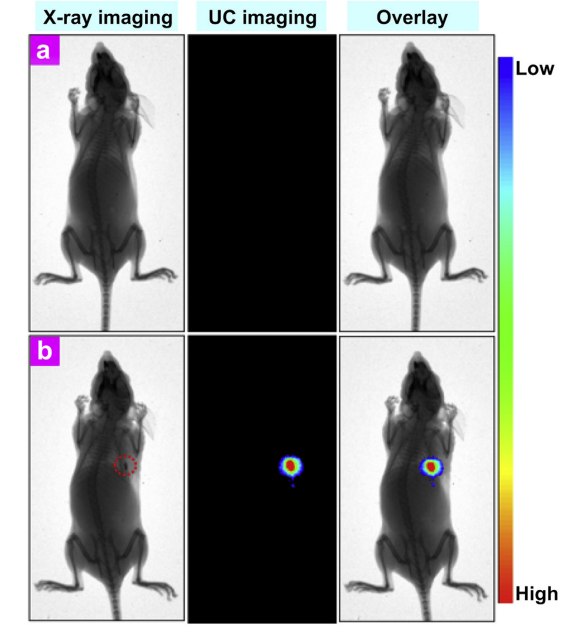
Figure 2: dual-modal X-ray and UC optical bioimaging of a nude mouse: (a) without injection of the ligand-free NaLuF4:Gd nanoparticles, (b) without injection of the ligand-free NaLuF4:Gd nanoparticles [2]
Computed Tomography is an epitome of biomedical imaging. Without surgery and pain-free, CT is widely used as a first tool to help doctors diagnose a patient, especially for fracture. By using contrast agents, CT’s pixel improves further for similar density or similar atomic structure areas. Rare-earth nano CAs can further improve general clinical CAs’ drawbacks such as toxicity and low biocompatibility and give more possibility to combine several biomedical imaging at the same time to support each other.
Eric Loewen
Eric Loewen graduated from the University of Illinois studying applied chemistry. His educational background gives him a broad base from which to approach many topics. He has been working with topics about advanced materials for over 5 years at Stanford Materials Corporation (SMC). His main purpose in writing these articles is to provide a free, yet quality resource for readers. He welcomes feedback on typos, errors, or differences in opinion that readers come across.

