Rare-earth elements (REE) consist of the lanthanides (atomic number 57 to 60) plus Scandium (Sc) and Yttrium (Y). They often appear together in minerals and have similar physical and chemical properties. Rare-earth element is one of the most important strategic resources in 21 century. With the research on rare earth chemistry, they have been widely used in agriculture, chemical industry, biomedicine, and military as high-performance permanent magnets, catalysts, alloys, and so on. This article will focus on rare earth application in bioimaging, mainly based on the principle of magnetic resonance imaging (MRI) and the use of REE nanoparticles.
Biomedical imaging becomes a very helpful tool for researchers to visualize and understand biological processes at the molecular level, and for physicians to depict the inside picture of a body for further diagnosis or treatment. Medical imaging needs to be accurate and clear to give physicians more chances to detect lesions and take proper treatment to increase the survival ratio. To achieve that, detection methods, imaging properties, and imaging procedures are 3 important aspects that need to be improved continuously to provide better biomedical imaging.
Magnetic Resonance Imaging (MRI), X-ray imaging, Computed Tomography (CT), and Optic imaging are the main detecting methods used in biomedical imaging. They have the following advantages:
However, biomedical imaging has difficulties in detecting very small tissues unless it grows larger. Cancerous tissue, for example, cannot be detected by biomedical imaging until the tissue grows 3 or 4cm. Bioimaging contrast agents play an important role to solve this problem. Let’s first get some basic knowledge about MRI working principles.
The basic principle for MRI is the detection of the intensity difference of every pixel or voxel caused by the magnetic field changes. All atoms have nuclear spin such as H, C, and O. As we all know, 60%~70% of the human body are water (H2O). In other words, hydrogen (H) is the most abundant atom in the human body. H is used as an employed atom in MRI process because of its large abundance and its second most sensitive atom characteristic.
Let’s take one proton in the body as an example to show how MRI works.
Without any magnetic field applied to a body, all protons may point up or down and finally they can cancel each other out. So initially, the total proton spins are 0.
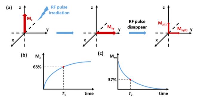
MRI scanner produces a strong magnetic field that makes the proton line up in one direction. As shown in the left image in Figure 1 a., the proton shows a total magnetization Mz in the z direction.

Figure 1: magnetic resonance imaging explanation. (a) The proton’s magnetization changes when RF pulse is applied and disappears. (b) &(c) Mz and Mxy change when RF pulse disappears [2].
Step 2
Then, Radio waves (RF pulse irradiation) sent from the MRI machine is applied to this proton. The proton’s magnetization will flip away from its z direction and point to the x,y direction. Mxy shows the excited state of the proton, as you cansee in the middle image in Figure 1a.
Step 3
Turn off the radio wave and the RF pulse disappears. Mz starts to appear and increases, while Mxy starts to disappear and decreases to 0. The proton can’t maintain the excited state, continuously going back to its original state and giving out radio signals. The MRI machine detects and stores this signal to create an image.
Figure 2.[3] may help you understand the process.

Figure 2. How Does an MRI Scan Work?
Figure 1 b & c shows Mz and Mxy changes as RF pulse disappears. T1 is the relaxation time required for Mz to recover to 63% and T2 is the relaxation time required for Mxy to decrease to 37% than its maximum value [2].
T1 and T2 values are mostly affected by tissues such as chemical structure and mobility. The signal intensity that is used to form MRI contrast is affected by the relaxation time, proton density and pulse sequence [2]. But at first, MRI is not a very sensitive machine to depict the differences between the normal and abnormal parts, magnetic contract agent (CA) is used to improve MRI’s image quality.
Nanoparticles are particles with sizes from 1 to 1000nm. Small size gives them a large surface area/volume, great tunability for multifunctional applications, and good compatibility with biological barriers [1]. There are still some requirements for nanoparticles used in bioimaging to improve functional specifies in the biological tissue and not to affect the biological tissue’s functionality [1].
The size of nanoparticles used in bioimaging range is from 10 to 150nm. In this range, nanoparticles can stay long enough to accumulate in the interesting area, so MRI can get better images. If the size is too large, it’ll take more time for nanoparticles to be cleaned up or even be trapped in the reticuloendothelial system which may cause serious toxicology issues [1].
Surface treatment is also important for nanoparticles because they will be exposed to the biological system. Surface coatings such as polyethylene glycol (PEG) polymers, poly(maleic anhydride-alt-1-octadecene) (PMAO) or silica are used to prevent unwanted interactions with the biological system and lower toxic effects [1]. The surface charge should be slightly positive or slightly negative or neutral to prevent detection by the immune system.
Last but not least, nanoparticle toxicity needs to be considered carefully. Not only because we don’t want the inappropriate nanoparticle as a CA is limited by our biological system, but also because we don’t want the nanoparticle furtherly hurt our bodies [1].
Rare-earth nanoparticles are ceramic nanoparticles made by oxides, sulfides, halides or oxysalts that contain lanthanide group ions (usually with +3 charged ions) [1]. Due to their 4f or 4f-5d orbital electrons, rare-earth nanoparticles show unique optical properties. Electrons in 5s and 5p orbitals give them unique spectroscopic and magnetic properties because they are shielded by electrons in 4f and 5d orbitals [1]. Gd3+ has 7 electrons evenly distributed in the 4f orbital. It has the highest unpaired electrons and shows good performance in increasing the signal intensity. Gd nanoparticles such as Gd2O3 decrease T1 time and enhance T1-weighted images especially brightening vascular structures and breakdown in the blood-brain barrier. GdF3 nanoparticles are more suitable as host matrixes for luminescent centers [1]. By giving different coatings such as 2-aminoethyl phosphate and poly(maleic anhydride-alt-1-octadicene), Gd nanoparticle shows better performance in decreasing T1 as a rare-earth CA.
Gd nanoparticles can also be used to decrease T2. The coating is the most important factor. Gd nanoparticles functionalized with aspartic-dextran polymers show a significant reduction in T2 [1]. D-glucuronic acid-coated ultra-small Gd2O3 nanoparticles also reduce T2 time.
For more information about nano-sized rare-earth element, you can provide your application information to our technical staff for advice. Stanford Advanced Materials (SAM) has rich experience in producing rare-earth elements products for more than 10 years.
Eric Loewen
Eric Loewen graduated from the University of Illinois studying applied chemistry. His educational background gives him a broad base from which to approach many topics. He has been working with topics about advanced materials for over 5 years at Stanford Materials Corporation (SMC). His main purpose in writing these articles is to provide a free, yet quality resource for readers. He welcomes feedback on typos, errors, or differences in opinion that readers come across.


